The needle is injected from the outside under the coracoid process of the scapula and moved between it and the head of the humeral bone 3-5cm deep backwards. The joint can be reached from the outside if the needle runs between the humeral process of the scapula and head of the humeral bone inside and downward. From behind the needle is introduced into the cavity formed by humeral process of the scapula and the back edge of the deltoid muscle, it then runs upward and forward, aiming at the coracoid, 4-5 cm deep.
Puncture of the elbow joint
A forearm is bent to make a right angle. The needle is then injected from outside between the external condyle of the humerus bone and the ulnar process and run to the joint cavity above the head of the radial bone. To perform the puncture of the upper volvulus of the joint the needle is introduced above the olecranon apex and run downward and forward.
Fig 27. Humeral Arthrocentesis
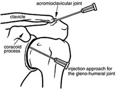
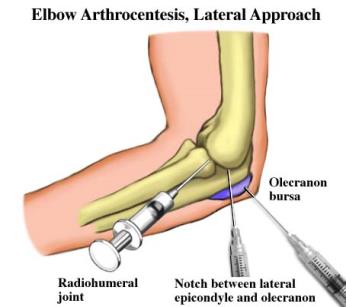
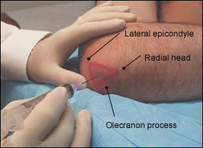
Fig 28. Puncture of elbow.
Puncture of the radiocarpal joint is performed on the dorsal surface of the hand. The projection of the joint corresponds to the line connecting the styloid process of the ulnar and the radial bones. The joint is punctured in the cross point of the connecting lines being the the continuation of the second metacarpal bone. The needle hits the joint between the long extensor tendons of the thumb and the extensor of the second finger.
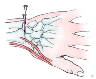
Fig 29. Puncture of a metacarpal joint.
Puncture of a hip joint
A needle is run in the frontal plane inside above the apex greater trochanter of the hip to cavity of acetabulum. The puncture can be performed from the front surface of the hip. The greater trochanter and the middle of poupart ligament are connected with a line its middle point corresponding to to the femoral head. The needle is introduced in this point at right angle to the skin and ran deep inside.
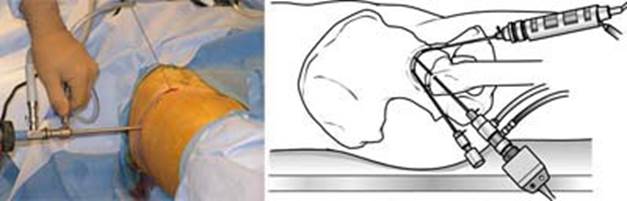
Fig 30. Puncture of a hip joint.
Puncture of the knee joint
The needle is introduced from the outer or inner side of the patella at the level of its middle height reaching between its back surface and front surface of the lower metaphysis of the hip. The top volvulus is punctured running the needle under the top point of the patella.
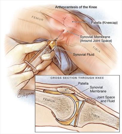
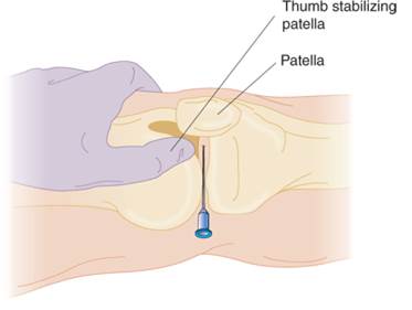
Fig 31. Puncture of the knee joint
Puncture of the ankle joint
The needle is injected 1, 5-2cm above and inward from the lateral malleolus apex. The needle is run backward horizontally with some down inclination and medially between lateral malleolus and long extensor tendon of the fingers. The joint cavity can be reached from the medial side. The needle is injected 1 cm higher than medial malleolus apex. The needle is than ran between the medial malleolus and anterior tibial muscle tendon.
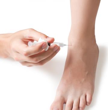
Fig 32. Puncture of the ankle joint
Дата: 2019-03-05, просмотров: 671.