ARTICULATIO GENU
The knee joint is the biggest and complexest of all the joints. It is formed by the femur, the tibia, the patella. On the superior articular surface of the tibia there is intra-articular cartilage or meniscus lateral and medial meniscus.
Anteriorly, the meniscus is connected by the intra-articular lig. Transversum genus. And also to the group of intra-articular ligaments are included the ligg. Cruciatum anterius et posterius. To the extra- articular ligaments includes: lig. Patella, lig. Collaterale tibiale, lig. Collaterale fibulare; posteriorly: lig. Popliteum arcuatum and lig. Popliteum obliquum.
In the region of the knee joint, there are numerous bursas that communicate and those that do not communicate with the joint cavity. They are bursa suprapatellaris, bursa prepatellaris subcutanea, bursa subfascialis prepatellaris, bursa subtendinea prepatellaris, and bursa infrapatellaris profunda.
The knee joint is supplied by the articular network, which is formed by aa. Genus superiores mediales et laterale, aa. Genus inferiores mediales et laterales, a. Genus media (a. Poplitea), a. Genus descendens (a. Femoralis), aa. Recurrentes tibiales anterior et posterior (a. Tibiales anterior).
FOSSA POPLITEA
The popliteal fossa is limited: medially above by m. Semitendinosus et m. Semimembranosus, laterally above by m. Biceps femoris, inferiorly by m. Gastrocnemius (caput laterale et caput mediale).
The layer-by layer structure of the region: skin, subcutaneous fatty layer, superficial fascia and the proper fascia (popliteal fascia). In the subcutaneous fatty layer passes: v. Saphena magna and n. saphenus medially, v. Saphena parva et n. cutaneous surae medialis laterally. N. cutaneous femoris posterior also pass through the sub cutaneous fatty layer.
The primary neuro-vascular bundle consists of n. tibialis (n. ischiadicus) superficially and laterally; v.poplitea deeply and medially. Laterally along the tendon of m. Biceps femoris passes the n. perneous communis (n. ischiadicus).
A. poplitea is the continuation of a. Femoralis. In the popliteal fossa, a. Poplitea is located on the bone itself, where it can be pressurised to stop bleeding.
Branches of a. Poplitea:
A) Which branch in the popliteal fossa:
1. Aa. Genus superiores lateralis et mediales.
2. Aa. Genus inferiores lateralis et mediales
3. Aa. Genus media
B) Terminal branches:
4. A. Tibiales anterior
5. A. Tibiales posterior
REGIO CRURIS
The crural region is limited superiorly by a horizontal line which passes through the tibial tuberositas and fibular capitulum; inferiorly by a horizontal line which passes through the lateral malleolus of the fibula and medial malleolus of the tibia.
The layer-by layer composition of the region: skin, subcutaneous fatty layer, superficial fascia, proper fascia (crural fascia).Through the subcutaneous fatty layer passes: v. Saphena magna and n. saphenus medially, v.saphena parva and n. cutaneous surae medialis laterally. And also in the subcutaneous fatty layer passes thye n. cutaneous surae lateralis, which in the lower third of the thigh, along with n. cutaneous surae medialis n. suralis and n. peroneus superficialis (n. peroneus communis).
The proper fascia of the crural region forms a septum, which divides the region into three fascial beds. Large neurovascular bundles pass through each of these fascial beds.
The anterior neurovascular bundle consists of a.v. tibialis anterior and n. peroneus profundus. A. Tibialis anterior (a. Poplitea) in the upper thirds of the thigh, passes between m. Tibialis anterior and m. Extensor digitorum longus, and still below lies between m. Tibialis anterior and m. Extensor hallucis longus. In the upper part, the nerve is located laterally and in the lower parts is located medial to the artery.
Branches of a. Tibialis anterior:
1. A. Recurrens tibiales posterior
2. A. Recurrens tibiales anterior.
3. Aa. Malleolares anteriores mediales et lateralis, which take part in the formation of the medial and the lateral malleolar network ( rete malleolare mediale et laterale).
In the anterio- lateral bed there are two canals:
1. Superior musculoperoneal canal is formed by the fibula and m. Peroneus longus. N. peroneus superficialis passes throught the canal.
2. Inferior musculoperoneal canal is formed by the fibula and m. Flexor hallucis longus. A.v. peronea passes through the canal.
The posterior neuro-vascular bundle consists of a.v. tibiales posterior and n. tibialis. A. Tibiales posterior (a. Poplitea) passes through the cruropopliteal canal. In the upper parts the foramen is limited by m. Soleus et caput laterale et mediale m. Gastrocnemius. The cruropopliteal canal is limited anteriorly by m. Tibiales posterior, and posteriorly by m. Soleus, laterally by m. Flexor hallucis longus, medially by m. Flexor digitorum longus.
The branches of a. Tibiales posterior:
1. A. Plantaris medialis.
2. A. Plantaris lateralis.
3. A. Peronea (fibularis)
REGIO MALLEOLUS MEDIALIS
The borders of the region correspond to that of the borders of malleolus medialis and Achille’s tendons. Through the subcutaneous fatty layer passes n. saphena magna and n. Saphenus. The malleolar canal is limited by the medial malleolus, retinaculum flexorum and the Achille’s tendon. Through the canal passes the tendons of m. Flexor digitorum longus, m. Tibiales posterior, m. Flexor hallucis longus, a.v.v. tibiales posterior and n. tibialis. The neuro-vascular bundle gets divided into 2 (a.v.v.n. plantares lateralis et mediales).
REGIO PEDIS
DORSUM PEDIS:
In the subcutaneous fatty layer is located the venous network of the dorsal pedis, and v. Saphena magna starts medially and v. Saphena parva starts laterally. The skin i the region is innervated by n. saphenus, n. cutaneus dorsalis pedis lateralis (n. suralis), n. peroneus superficialis et profundus.
The primary artery of the region is a. Dorsalis pedis.
Branches of a. Dorsalis pedis:
1. Aa. Tarseae mediales
2. A. Tarsea lateralis
3. A. Arcuata. From this artery branches out three aa. Metatarsea dorsales, each of which gets divided into two aa. Digitales dorsales communis, which continues as aa. Digitales dorsales propria;
4. A. Metatarsea dorsalis prima
5. Ramus plantaris profundus.
PLANTA PEDIS
Regio planta pedis is supplied by aa. Plantares mediales et laterales. A. Plantaris medialis is located in the sulcus plantaris medialis. The larger a. Plantaris lateralis passes into the sulcus plantaris lateralis and takes part in the formation of arcus plantaris.
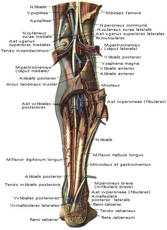
Fig. 12. Topography of region fossa poplitea et regio cruris posterior.
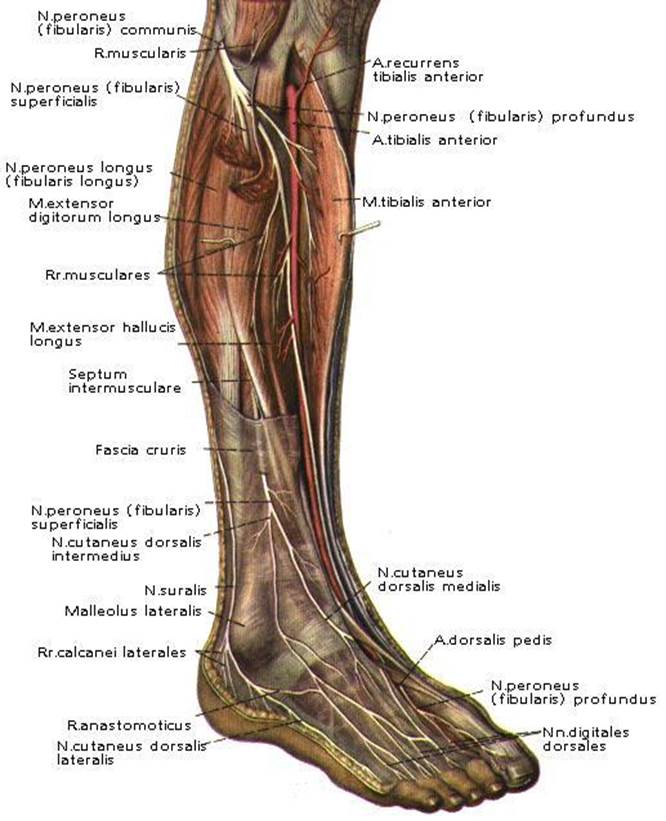
Fig. 13. Topography of region cruris anterior.
ЗАНЯТИЕ № 5.
ТОПОГРАФИЯ ARTICULATIO GENU, FOSSA POPLITEA, REGIO CRURIS, REGIO MALLEOLUS MEDIALIS, REGIO PEDIS.
ARTICULATIO GENU .
Art. genu, является самым большим и сложным из всех суставов. В его образовании принимают участие: os femor, os tibia, patella. На facies articularis superior os tibia имеются внутрисуставные хрящи, или мениски – meniscus lateralis et medialis. Спереди мениски соединены внутрисуставной связкой lig.
transversum genus. Также к внутрисуставным связкам относятся ligg. cruciatum anterius et posterius. К внесуставным связкам относятся: передние – lig. patellae, lig. collaterale tibiale, lig. collaterale fibulare; задние – lig. рорliteum arcuatum и lig. popliteum obliquum.
В region articulatio genu имеется ряд сумок сообщающихся и не сообщающихся с полостью сустава. Это bursa suprapatellaris, bursa prepatellaris subcutanea, bursa subfascialis prepatellaris, bursa subtendinea prepatellaris, bursa infrapatellaris profunda.
Art. genu кровоснабжается rete articulare, которая образована aa. genus
superiores medialis et lateralis, aa. genus inferiores medialis et lateralis, a. genus media (a. poplitea), a. genus descendens (a. femoralis), aa. recurrentes tibiales anterior et posterior (a. tibialis anterior).
FOSSA POPLITEA.
Fossa poplitea ограничена сверху медиально – m. semitendinosus et m. semimembranosus, сверху латерально – m. biceps femoris, снизу – m. gastrocnemius (caput laterale et caput mediale).
Послойное строение – кожа, подкожная жировая клетчатка, поверхностная фасция, собственная фасция (fascia poplitea). В подкожной жировой клетчатке проходят: медиально – v. saphena magna et n. saphenus, латерально – v. saphena parva et n. cutaneus surae medialis. Также в подкожной жировой клетчатке проходят n. cutaneus surae lateralis et n. cutaneus femoris posterior.
Основным сосудисто-нервным пучком является: поверхностно латерально – n. tibialis (n. ischiadicus); глубже, медиально – v. poplitea; еще глубже и медиально – a. poplitea. Латерально, вдоль tendo m. biceps femoris проходит n. peroneus communis (n. ischiadicus).
A. poplitea, продолжение a. femoralis. В fossa poplitea a. poplitea располагается на самой кости, где ее можно пережать.
Ветви a . poplitea :
А) Отходящие в fossa poplitea.
1. Aa. genus superiores lateralis et medialis.
2. Aa. genus inferiores lateralis et medialis.
3. A. genus media.
В) Конечные ветви.
4. A. tibialis anterior.
5. A. tibialis posterior.
REGIO CRURIS.
Regio cruris ограничена сверху горизонтальной линией, проходящей через tuberositas tibia et capitulum fibula; снизу горизонтальной линией, проходящей через malleolus lateralis os fibula et medialis os tibia.
Послойное строение – кожа, подкожная жировая клетчатка, поверхностная фасция, собственная фасция (fascia cruris). В подкожной жировой клетчатке проходят: медиально – v. saphena magna et n. saphenus, латерально – v. saphena parva et n. cutaneus surae medialis. Также в подкожной жировой клетчатке проходят n. cutaneus surae lateralis, образующий в нижней трети голени вместе с n. cutaneus surae medialis n. suralis et n. peroneus superficialis (n. peroneus communis).
Собственная фасция голени дает перегородки, которые разделяют голень на три фасциальных ложа. В каждом фасциальном ложе проходят крупные сосудисто-нервные пучки.
Передний сосудисто-нервный пучок состоит из a.v. tibialis anterior et n. peroneus profundus. A. tibialis anterior (a. poplitea) в верхней трети голени, проходит между m. tibialis anterior и m. eхtensor digitorum longus, а ниже лежит между m. tibialis anterior и m. eхtensor halluсis longus. Вверху нерв расположен латерально, а внизу медиально артерии.
Ветви a . tibialis anterior :
1. A. reсurrens tibialis posterior.
2. A. reсurrens tibialis anterior.
3. Aa. malleolares anteriores medialis et lateralis, участвуют в образовании retе malleolaгe mediale et lateгale.
В передне-латеральном ложе голени имеется два канала.
Canalis musculoperoneus superior образован os fibula et m. peroneus longus. В нем проходит n. peroneus superficialis.
Canalis musculoperoneus inferior образован os fibula et m. flexor hallucis longus. В нем проходит a.v. peronea.
Задний сосудисто-нервный пучок состоит из a.v. tibialis posterior et n. tibialis. A. tibialis posterior (a.poplitea) идет в сanalis сruropopliteus. Верхнее отверстие канала ограничено m. soleus et capus laterale et mediale m. gastrocnemius. Canalis сruropopliteus ограничен спереди – m. tibialis posterior, сзади – m. soleus, латерально – m. fleхor halluсis longus, медиально – m. flexor digitorum longus.
Дата: 2019-03-05, просмотров: 1051.