AXILLARY FOSSA AND THE BRACHIAL REGION.
DELTOID REGION
The deltoid region indicates the localisation of the deltoid muscles and the humeral articulation. The proper fascia of the deltoid region forms the vagina for the m. Deltoideus. The processes penetrating through the muscles and divide it into fascicles extends from the fascia to the muscles. Between m.deltoideus and the humeral bone is located the cellulose space spatium subdeltoideum, through which passes n.axillaris and a.Circumflexa humeri posterior.
HUMERAL ARTICULATION
Caput humeri and cavitas glenoidealis scapulae form the humeral articulation.
The articulation is held in place with the help of 5 ligaments: lig. Coracoacromiale, lig. Coracohumerale, lig. Glenohumerale superioris, lig. Glenohumerale inferioris, lig. Glenohumerale media.
The synovial bursa is located near the joint. Two of these bursas do not communicate with the joint cavity: bursa subdeltoidea, bursa subacromialis. Bursa subscapularis and bursa subcoracoidea communicate with the joint cavity. The joint capsule is attached to the surgical neck of the humeral bone. The cavity of the humeral joint is expanded with the help of three rotations: recessus axillaris, recessus subscapularis, and recessus intertubercularis.
SUBCLAVIAN REGION
The layer-by-layer structure of this region is represented by the skin, subcutaneous fatty tissue, superficial fascia, proper fascia, m.pectoralis major et minor.
There are three triangles in the subclavian region:
1. Trigonum claviculopectorale [bordered superiorly by the clavicle, medially by the sternum, laterally downwards by m.pectoralis minor]
2. Trigonum pectorale [corresponds to the borders of m.pectoralis minor]
3. Trigonum subpectorale [corresponds to the borders of fossa axillaris]
The important neurovascular fascicle in the subclavian region is a.v. subclavia, plexus brachialis. The continuity of a. Subclavia is a. Axiallaris, which in turn continues as a. Brachialis.
The proximal borders of the trunk of a.axillary is the lateral border of the first rib, and the distal border is the inferior border of m. Teres major (place of beginning of a. Brachialis)
A. axillaris is located in the axillary cavity medial to the humeral joint cavity and the humeral bone; medially and anteriorly to the artery are located v. Axillaris and from all the three sides is surrounded by the trunk of plexus brachialis; inferiorly the neuro vascular fascicle is covered by skin, fascia and mass of fatty tissues, which consist of the lymph nodes.
A. axillaris gets divided into three sections:
1) From the clavicle, medial border of the sternum upto the superior border of m. Pectoralis minor (trigonum claviculopectorale)
2) Behind m. Pectoralis minor (trigonum pectorale)
3) From the inferior border of m. Pectoralis minor upto the inferior border of m. Pectoralis major (trigonum subpectorale)
The branches of a. Axillaris in the claviculopectoral triangle are:
1. A.thoracic superior gets divided in m. Subclavius, m. Pectoralis major et minor, m. Serratus anterior superior, m. Intercostalis
2. A. Thoracoacromialis takes part in the nourishment of the humeral articulation, m. Deltoideus, m.pectoralis major and minor.
The branches of a. Axillarisin the pectoral triangle are:
3. A. Thoracica lateralis descends down along the lateral wall of the thorax and gives branches to the mammary glands and the surrounding muscles.
The branches of a. Axillaris in the subpectoral triangle or in the axillary fossa are:
4. A. Subscapularis, which is the largest branch of a. Axillary, starts from the inferior border m. Subscapularis, descends along this muscle and divides into two trunks:
a) A. Circumflexa scapulae passes through the trilateral foramen onto the dorsal surface of the scapula
b) A. Thoracodorsalis passes along the posterior surface of the thorax
5. A. Circumflexa humeri posterior passes posteriorly, into the quadrilateral foramen, curves around the surgical neck of the humeral bone and is supplies m. Deltoideus.
6. A. Circumflexa humeri anterior passes in the lateral direction, circumflexes around the the surgical neck of the humeral bone anteriorly, and anastomizes with a. Circumflexa humeri posterior.
AXILLARY FOSSA
The axillary fossa is cone shaped. It is bordered: anteriorl by the inferior border of m. Pectoralis major, posteriorly by m. Latissimus dorsi, medially by the line that joins the borders of these muscles on the thorax, laterally by the line that joins borders of these muscles on the humeral bone.
The walls of the axillary fossa are: anterior- m.pectoralis major et minor; posterior- m.latissimus dorsi, m. Teres major et minor, m.subscapularis; lateral- m. Corachobrachialis; medial- m. Serratus anterior superior.
On the posterior wall of the axiallary fossa are located two foramens: trialteral and quadrialateral. The trilateral foramen is bordered: superiorly by m. Teres minor, m. Subscapularis; inferiorly by m. latissimus dorsi, m. Teres major; laterally bycaput longum m. Triceps brachii. A. Circumflexa scapula passes through this foramen.
The quadrialteral foramen is bordered: superiorly by m. Teres minor, m. Subscapularis; inferiorly m.latissimus dorsi, m. Teres major; laterally by the humeral bone; medially by the caput longum m. Triceps brachii. N. axillaris and a. circumflexa humeri posterior pass through this foramen.
The important neuro-vascular fascicle in this fossa consist of the superficially located v. Axillaris, laterally and deeply located a. Axillaris, which is surrounded by three fascicles of the brachial plexus (lateral, medial and posterior). From the fascicle of the brachial plexus begins the nerves in the axillary fossa. From the posterior bundle starts n. radialis, n. axillaris; from the medial bundle starts medial radix of n. medianus, n. ulnaris, n. cutaneus brachii medialis, n. cutaneus antebrachii medialis; from the lateral bundle starts the lateral radix of n. medianus, n. musculocutaneus.
In the subcutaneous fatty layer are located 5 groups of lymph nodes: nodi. Lymphatici axillaris lateralis, nodi lymphatici axillaris medialis (pectoralis), nodi lymphatici axillaris centralis, nodi lymphatici axillaris subscapularis, nosi lymphatici axillaris apicalis
BRACHIAL REGION
This region is limited: superiorly by the line, that joins the borders of m. Pectoralis major and m. Latissimus dorsi; inferiorly by the line that passes at a level of two fingers above the humeral epicondilus. The skin, subcutaneous fatty tissues, superfiscial and proper fascia represent the layers of this region. The proper fascia surrounds the muscles and the neuro-vascular bundles of the arm.
Through the subcutaneous fatty tissues pass the superfiscial veins laterally, v. Cephalica passes through the lateral bicipital sulcus; and medial to the medial bicipital sulcus passes v. Basilica together with n. cutaneous antebrachii medialis. The skin on the arm is innervated b n. cutaneous brachii lateralis, medialis et posterior.
The important neuro-vascular bundles in the arm are a.v.v. brachiales and n. medianus. The bundle passes through the medial bicipital sulcus. In the upper third of the arm, the nerve is located lateral to the artery, and in the middle third on the artery, and in the lower third medially from the artery. The second neuro-vascular bundle in this region are the a.v.v collateralis ulnaris superior and n. ulnaris, which in the middle third passes through the bed formed by m. Triceps brachii. The third neuro-vascular bundle in the region consists of a.v.v profunda brachii and n. radialis that are located in the posterior surface of the arm and passes through the humero-muscular canal (spiral canal, canal of n. radialis). M. Triceps brachii and the humerus bone form the canal. The branches of a. Brachialis- a. Collateralis ulnaris superior and inferior, and a. Profunda brachii and its branches a. Collateralis media and a. Collateralis radialis accomplish the collateral blood supply to the arm.
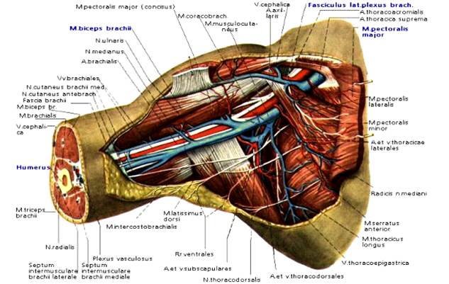
Fig. 6. Fossa axillaris.
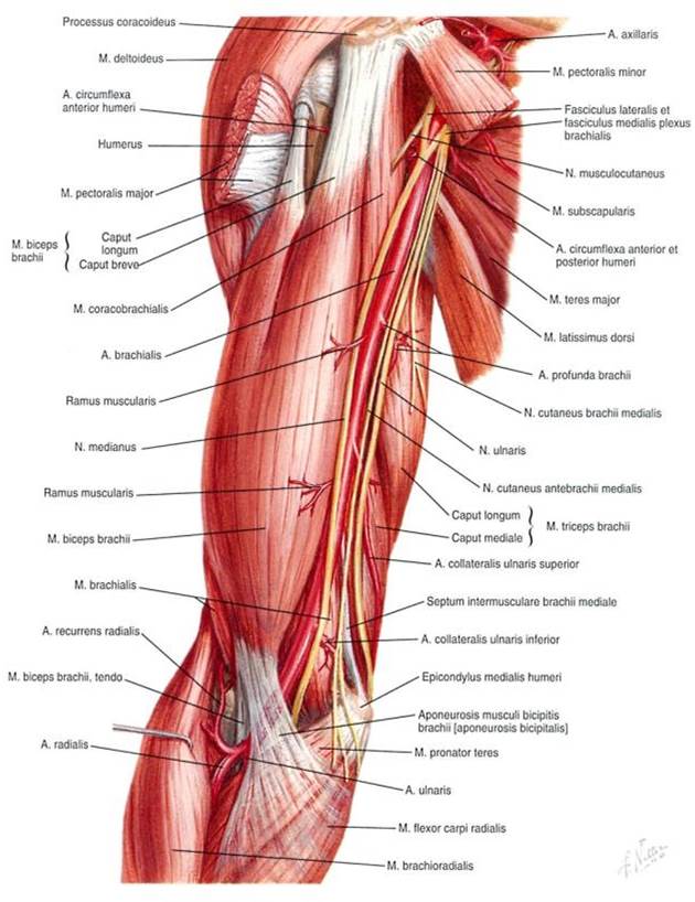
Fig. 7. Regio brachii.
ЗАНЯТИЕ № 2.
Дата: 2019-03-05, просмотров: 963.