(CRANIOECTOMIA)
Indicators: tumors of brain, cerebral hernias, edema of brain, cyst of brain and others.
Position of patient depends upon the location of pathological process and selection of surgical approach, projection of which is determined on cranial surface with the help of Cronleyn-Brjusova’s scheme.
Techniques of operation include:
1- arch section of skin up to periosteum is performed. Aponeurosal-cutaneous flap is separated and turned downwards. Stopping of bleeding. Flap is closed by napkin and then attached with operation linen.
2- periosteum is incised by arched section, at a distance of 1-2 cm from the margin of wound. It is then removed by raspatory on the direction opposite of the incision in order to form osteoperiosteal flap.
3- Along the line of incision of periosteum, 4-5 miller holes are drilled and bone between them is sawed through by wire saw with the help of guides.
4- Osteoperiosteal flap is cracked between 1st and 5th hole and thrown away below, preserving its connection with periosteum.
5- Dura mater is incised by arch section and flap is turned towards the direction of saggital sinus.
6- Necessary manipulations are done in cranial cavity. Operation is performed carefully without excessive brain injury of brain, because there is the possibility of edema of cerebral tissue during postoperative period.
7- Dura mater is sutured with interrupted or continuous sutures.
8- Layer by layer suturing of wound.
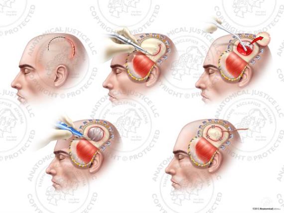
Fig 63. Craniotomy.
MASTOIDOTOMY (ANTROTOMIA)
Indicators: acute purulent inflammation of cells of processus mastoideus (mastoiditis), chronic inflammation of middle ear.
Techniques of operation:
1- arched incision of skin and subject tissues together with periosteum, on 1 cm behind the line of attachment of auricle.
2- Periosteum is removed aside by raspatory and external surface of processus mastoideus is uncovered. Within Shipo triangle, cortical layer is removed with the help of gouge and hammer. Trepanation aperture is continuously dilated to open widely the main cell of the processus mastoideus and the adjoining cells to it, containing pus- forming one big cavity. While working with cell, one should remember the location of facial nerve, sigmoid venous sinus and middle cranial fossa, which may get injured by instruments or debris. Drainage is inserted in the lower angle of the wound and 2-3 silk sutures are placed on skin.
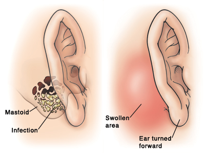
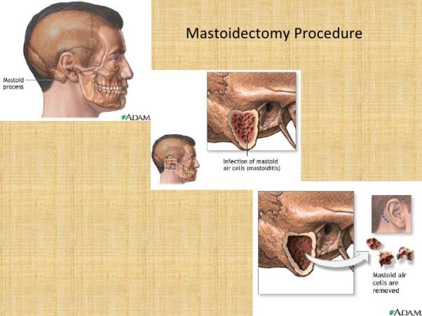
Fig 64. Mastoidectomy.
Дата: 2019-03-05, просмотров: 674.