AND THE REGION OF PALMA MANUS
FOSSA CUBITI
The cubital fossa is limited above by m. Brachioradialis laterally, by m. Biceps brachii medially, and below by m. Brachioradialis laterally, by m. Pronator teres medially. The layers of this region are: skin, subcutaneous fatty layer, the proper fascia, the surrounding neuro-vascular bundles and the muscles of the region.
In the subcutaneous fatty layer is located the superficial veins and the cutaneous nerves: laterally - v. Cephalica, n. cutaneous antebrachii lateralis (enters into the cubitalfossa in between m. Biceps brachii and m. Brachialis); medially - v. Basilica and n. cutaneouss antebrachii medialis. The veins form two different anastomoses between themselves:
1. < N > shaped (v. Mediana cubiti)
2. < M > shaped (v. Mediana cephalica, v. Mediana basilica, v. mediana antebrachii)
Through the cubital fossa passes two neuro-vascular bundles. Laterally - n. radialis and a.v. collateralis radialis. The nerve leaves the fossa in between m. Brachialis and m. Brachioradialis. It branches into superficial ramus of n. radialis, which goes along the anterior surface of the forearm and the deep ramus of n. radialis, which passes through the supinator canal on the posterior surface of the forearm. The medial neuro-vascular bundle is formed by a.v.v brachialis and n.nmedianus. The artery passes medially from m. Biceps brachii, and the nerve passes 0,5-1,0 cm medially from the artery.
On the posterior surface of the cubital region is located the osteofibrous canal, which is formed by the medial epicondle of the humerus and the olecranon of the ulna. Through the canal passes n. ulnaris and a.v. collateralis ulnaris superior.
CUBITAL ARTICULATION
The cubital articulation is formed by 3 bones: The humerus, the ulna and the radius.
The joint is strengthened by 3 ligaments: lig. Anulare radii, lig. Collaterale ulnare, lig. Collaterale radiale. There are 2 weak spots in the capsule of the cubital articulation: They are the sacciform recessus and the posterio-superior area of the capsule.
In the region of the cubital articulation, there is a network formed bya. Recurrens ulnaris (a. Ulnaris), the branches of which anastomoses with a. Collateralis ulnares superior et inferior (a. Brachialis). A. Recurrens radialis forms ananstomoses with a. Collateralis radialis et media (a. Profunda brachii).
ANTEBRACHIAL REGION
The antebrachial region is limited: superiorly by 2 transverse fingers below the line, which joins the medial and lateral epicondyle of the humerus, inferiorly by the line that joins the styloideus process of the radius and ulna.
The layer-by-layer composition of this region: skin, subcutaneous fatty layer, superficial and the proper fascias.
Through the subcutaneous fatty layer passes v. Cephalica et n. Cutaneous antebrachii lateralis; v. Basilica et n. cutaneous antebrachii medialis.
The muscles of the anterior surface of the forearm are reapresented by four layers:
1 layer- m. Palmares longus, m. Pronator teres, m. Brachioradialis, m. Flexor carpi radialis. M. Flexor carpi ulnaris.
2 layer- m. Flexor digitorum superficialis
3 layer- m. Flexor digitorum profundus et m. Pollicis longus
4 layer- m. Pronator quadratus.
In the lower third of the forearm is located the cellulose space of Pirogov, which is limited by the 3rd and 4th layers of muscles.
A. Brachialis in the cubital fossa gets divided into two branches: a. Ulnaris, a.radialis
A. ULNARIS is the largest branch of a. Brachialis. In the upper third of the forearm the artery passes under m. Pronator teres. In the middle and lower thirds of the forearm a. Ulnaris and n. ulnaris pass in between m. Flexor digitorum superficialis and m. Flexor carpi ulnaris (forms the medial neuro-vascular bundle of the forearm). In the radial side of the pisform bone, a. Ulnaris and n. ulnaris passes into the canalis carpi ularis (interaponeurotic space)
The brances of a. Ulnaris:
1. a. Recurrens ulnaris
2. a. Interrossea communis goes to interosseal membrane and gets divided into 2 branches:
a) a. Interossea anterior passes along the anterior surface of the interosseal membrane, and gives a. Mediana.
b) a. Interossea posterior passes along the posterior surface of the forearm and together with the deep ramus of n. radialis forms the posterior neuro-vascular bundle of the forearm.
3. Ramus carpeus palmaris
4. Ramus carpeus dorsalis
5. Ramus palmaris profundus of a. Ulnaris forms anastomoses with a. Radialis and forms arcus palmaris profundus, which is located in between the tendon of m. Flexor digitorum and metacarpal bone.
A.v.v. Mediana and n. medianus forms the middle neuro-vascular bundle of the forearm. The middle third of the forearm passes between m. Flexor digitorum superficialis et profundus. In the lower thirds of the forearm, they pass between m. Flexor digitorum superficialis and m. Flexor carpi radialis.
A.v.v. interossea anterior and n. interosssea anterior forms the deep neuro vascular bundle of the forearm.
A. RADIALIS goes into the radia sulcus along with n. radialis in between m. Brachioradialis and flexor carpi radialis.
The branches of a. Radialis:
1. a. Recurrens radialis
2. Rami musculares (to the adjacent muscles)
3. Ramus carpeus
4. Ramus palmaris superficialis of a. Radialis anastomoses with ramus carpeus palmaris a. Ulnaris and forms arcus palmaris superficialis ( which is located in between the tendons of the flexor digitorum and the aponeurosis palmaris).
5. Ramus carpeus dorsalis
6. a. Metacarpea dorsalis prima
7. a. Princeps pollicis
REGIO PALMA MANUS
The layer-by-layer structure of the region: skin, subcutaneous fatty tissue, superficial fascia, proper fascia (aponeurosis palmaris). The proper fascia gives a septum, which divides Palma manus into 3 fascial beds: lateral (thenar), medial (hypothenar) and middle. In each of these beds are located the muscles, tendons, vessels, nerves, cellulose spaces. The muscles of the thenar are m. Abductor pollicis brevis, m. Opponens pollicis, m. Flexor pollicis brevis. Muscles of the hypothenar are mm. Flexor abductor opponens digiti minimi.
There are 4 cellulose spaces in the palm, where in case of pus formation, phlegmons are formed and are referred to as: thenar, hypothenar, sub aponeurotic, subtendonic, comissural.
Synovial vaginas of the tendons of the flexors of the fingers have a speciality. The synovial vagina of the first and the fifth fingers start from the region of the base of the distal phalange and ends in the lower third of the forearm in the cellulose space of Pirogov, forming the radial and ulnar synovial bags. The synovial vagina of the 2, 3, 4 th fingers starts from the region of the base of the distal phalange and ends in the region of the head of the carpal bones.
The wrist is supplied by the superficial and the deep arterial arches.
The palmar side of the wrist and the fingers are innervated by n. medianus et n. ulnaris. The dorsal side of the wrist and the fingers are innervated by n. radialis and n. ulnaris.
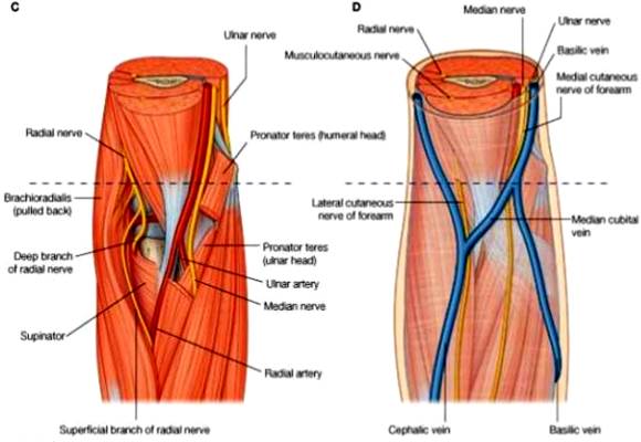
Fig. 8. FOSSA CUBITI, REGIO ANTEBRACHII, REGIO PALMA MANUS.
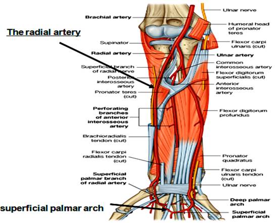
Fig. 9. FOSSA CUBITI, REGIO ANTEBRACHII, REGIO PALMA MANUS.
ЗАНЯТИЕ № 3.
Дата: 2019-03-05, просмотров: 419.